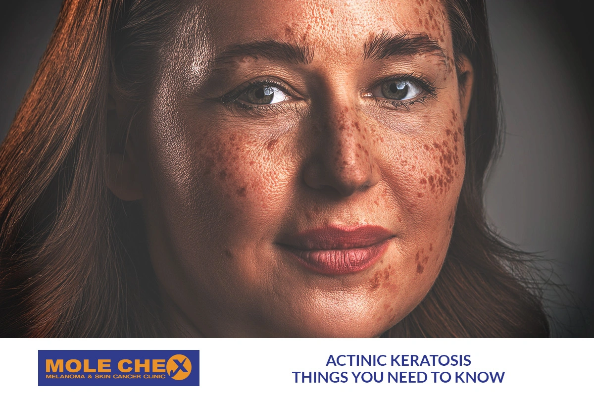
Not every rough, scaly patch on the skin is harmless. Some spots develop after years of ultraviolet exposure and may represent actinic keratosis, a precancerous skin condition that deserves professional attention. Knowing when to consult actinic keratosis specialists can help you recognise concerning changes, receive an accurate diagnosis, and reduce your risk of developing skin cancer.
Actinic keratosis commonly appears on sun-exposed areas, including the face, ears, scalp, neck, forearms, and backs of the hands. Early lesions can be subtle, making them easy to overlook. Although many remain noncancerous, some can progress to squamous cell carcinoma, a common type of skin cancer.
Understanding warning signs, available treatments, and appropriate follow-up can help you make informed decisions about your skin health.
What Is Actinic Keratosis?
Actinic keratosis, sometimes called solar keratosis, develops when ultraviolet radiation damages skin cells over time. Repeated exposure to sunlight or artificial tanning sources can cause abnormal changes in keratinocytes, the cells that form the outer skin layer.
The condition often develops gradually. A lesion may initially feel rough before becoming visibly scaly, crusted, or discoloured. Some spots are easier to feel than see, particularly when they resemble sandpaper.
People with lighter skin, a history of sunburn, extensive outdoor exposure, or previous skin cancer generally face a higher risk. However, actinic keratosis can occur in people with different skin tones.
These lesions are not contagious. Their presence indicates accumulated sun damage and provides an opportunity to assess skin health, discuss treatment, and improve sun protection.
What Do Suspicious Skin Spots Look Like?
Actinic keratosis does not have one universal appearance. Lesions vary in colour, thickness, size, and texture. Some are only a few millimetres wide, while others form larger patches.
Common characteristics include:
- Rough or sandpaper-like texture.
- Dry, flaky, or scaly surfaces.
- Pink, red, brown, tan, or skin-coloured patches.
- Small crusts that repeatedly return.
- Mild itching, tenderness, or burning.
- Persistent spots on frequently exposed areas.
The lips may also develop sun-related damage, particularly the lower lip. Persistent dryness, scaling, cracking, or a rough patch should not automatically be attributed to ordinary chapping.
A lesion can look relatively harmless while still requiring examination. Therefore, appearance alone cannot reliably determine whether a spot is actinic keratosis, another skin condition, or cancer.
When Should You See a Skin Specialist?
Arrange an appointment with a dermatologist if a rough or scaly spot persists, returns repeatedly, or develops without an obvious explanation. Early assessment is particularly important when a lesion appears on a sun-exposed area or you have previously experienced skin cancer.
Seek prompt medical assessment if you notice:
- Rapid growth or a sudden change in appearance.
- Bleeding without an obvious injury.
- An open sore that does not heal.
- Increasing pain, tenderness, or inflammation.
- A thick, raised, or unusually hard lesion.
- Repeated crusting after the surface appears to heal.
- A new growth that looks different from surrounding spots.
These changes do not automatically mean cancer is present. However, they can indicate a lesion that needs closer investigation.
Do not wait for a spot to become painful before seeking advice. Some important skin changes cause little discomfort during their early stages.
Who Treats Actinic Keratosis?
Dermatologists are doctors trained to diagnose and treat diseases affecting the skin, hair, and nails. They are generally the most appropriate professionals for assessing suspicious lesions and determining whether treatment or biopsy is necessary.
Experienced actinic keratosis specialists can distinguish many typical lesions from similar conditions, including eczema, psoriasis, seborrhoeic keratoses, warts, and certain skin cancers.
Depending on the findings, your care may involve a general dermatologist, a dermatologist with a skin cancer focus, or a specialist in dermatologic surgery.
A primary care doctor can also examine an unfamiliar lesion and arrange referral when necessary. If a spot appears particularly concerning, ask whether a prompt dermatology appointment is appropriate.
When choosing a clinician, consider their experience with precancerous lesions, access to biopsy services, explanation of treatment options, and arrangements for follow-up.
How Is Actinic Keratosis Diagnosed?
Visual Examination
Diagnosis often begins with a medical history and careful examination of the affected skin. The clinician may ask about sun exposure, previous skin cancers, medications, immune suppression, and changes in the lesion.
A dermatoscope, a handheld device that magnifies skin structures, may help the clinician examine features that are difficult to see with the naked eye.
When Is a Biopsy Needed?
A biopsy involves removing a small sample of tissue for laboratory examination. It may be recommended when the diagnosis is uncertain or a lesion has concerning features.
Biopsy becomes particularly important when a spot is thick, painful, rapidly growing, persistently ulcerated, or resistant to expected treatment.
Not every actinic keratosis requires biopsy. The decision depends on its appearance, clinical history, and response to treatment.
Accurate diagnosis matters because a suspected precancerous lesion may occasionally be squamous cell carcinoma or another condition requiring a different approach.
Treatment Options Recommended by Dermatologists
Treatment depends on the number of lesions, their location, thickness, previous treatments, and the patient’s overall skin cancer risk.
Cryotherapy
Cryotherapy uses extreme cold, usually liquid nitrogen, to destroy individual lesions. It is commonly used for a limited number of actinic keratoses.
The treated area may sting, blister, crust, and temporarily change colour. Healing time varies according to lesion size, location, and individual skin characteristics.
Prescription Creams and Gels
Certain prescription medicines treat visible lesions or broader areas of sun-damaged skin. Options may include fluorouracil, imiquimod, or other appropriate topical treatments.
These medicines can cause redness, irritation, crusting, and temporary inflammation. The expected reaction and treatment duration depend on the specific product and regimen.
Follow the prescribing clinician’s instructions carefully. Do not apply prescription products to suspicious lesions without appropriate medical guidance.
Photodynamic Therapy
Photodynamic therapy uses a light-sensitive medication followed by a specific light source to destroy abnormal cells. It may be suitable when several lesions affect one area.
Temporary redness, swelling, discomfort, and sensitivity to light can occur. Your clinician will explain preparation, aftercare, and sun avoidance requirements.
Curettage and Other Procedures
Curettage involves scraping away abnormal tissue using a specialised instrument. It may be considered for selected thicker lesions, sometimes alongside another procedure.
Other treatments may be appropriate according to the diagnosis and clinical circumstances. Your dermatologist can explain likely benefits, side effects, healing expectations, and recurrence risks.
Can Natural Approaches Help?
People often prefer gentle skincare and lifestyle changes, especially when dealing with recurring sun damage. However, natural approaches should complement, not replace, medical assessment and proven treatment.
A Natural Treatment for Actinic Keratosis should not be assumed to remove abnormal cells simply because an ingredient is plant-derived. No home remedy has been established as a reliable substitute for appropriate clinical care.
Avoid applying acidic household substances, essential oils, abrasive scrubs, or untested preparations to suspicious spots. These can irritate skin, cause burns, or delay diagnosis.
Gentle cleansing, fragrance-free moisturising products, protective clothing, and consistent sun protection can support the skin barrier. Ask your dermatologist before using any product directly on a diagnosed lesion, particularly if it is bleeding, inflamed, or undergoing treatment.
What About Herbal and Home Remedies?
Interest in botanical skincare is understandable, but evidence for treating precancerous lesions with herbs remains limited. A product’s natural origin does not establish its safety or effectiveness.
Some Natural Remedies for Actinic Keratosis are promoted online without convincing clinical evidence. They should never be used to postpone assessment of a persistent, changing, or suspicious spot.
If you use Herbal Care Products for general skincare, check the ingredient list and discontinue use if irritation develops. Avoid products that promise to cure precancerous lesions without medical treatment or reliable evidence.
Discuss supplements and topical preparations with your healthcare professional, particularly if you take medication, have allergies, or have sensitive skin. Evidence-based treatment and appropriate monitoring remain essential.
Why Sun Protection Matters
Sun protection helps reduce additional ultraviolet damage and may lower the likelihood of developing further actinic keratoses. It remains important after treatment because removing existing lesions does not reverse every change caused by earlier exposure.
Practical measures include:
- Apply broad-spectrum sunscreen with SPF 30 or higher.
- Reapply sunscreen at least every two hours outdoors and after swimming or heavy sweating.
- Wear a broad-brimmed hat and tightly woven protective clothing.
- Seek shade when sunlight is strongest.
- Avoid indoor tanning beds.
- Use a lip balm with broad-spectrum sun protection.
Remember to protect the ears, scalp, neck, hands, and other exposed areas. People with thinning hair may need sunscreen or protective headwear for the scalp.
No sunscreen offers complete protection. Combining several protective measures is more reliable than depending on one product alone.
What Happens If Actinic Keratosis Is Ignored?
Some lesions remain stable for long periods, while others disappear or return. Others may become thicker or develop into squamous cell carcinoma. It is not possible to predict an individual lesion’s behaviour with certainty from appearance alone.
Leaving a suspicious spot unchecked can delay diagnosis of skin cancer or another condition requiring treatment. This does not mean every untreated lesion will become malignant; rather, the uncertainty makes appropriate evaluation important.
A clinician can assess which lesions need treatment, which require biopsy, and how frequently your skin should be checked.
People with numerous lesions or a history of skin cancer may benefit from a personalised surveillance plan. Regular appointments also provide an opportunity to discuss new spots and reinforce sun protection habits.
What to Expect During Your Appointment
Preparing for a dermatology visit can make the consultation more useful. Write down when you first noticed the lesion, whether it has changed, and any symptoms you have experienced.
Bring a list of medicines, skincare products, and relevant medical history. If the spot changes intermittently, photographs taken over time may help demonstrate its development, although they cannot replace an examination.
During the appointment, actinic keratosis specialists may examine the lesion and surrounding skin, recommend a biopsy, or discuss immediate treatment. You should understand the reason for the proposed approach and how to care for the area afterwards.
Useful questions include:
- What is the most likely diagnosis?
- Is a biopsy necessary?
- Which treatment suits this lesion?
- What side effects or healing changes should I expect?
- When should I return for reassessment?
- What changes should prompt an earlier appointment?
Do not hesitate to ask for clarification if a treatment plan or aftercare instruction is unclear.
How Often Should Your Skin Be Checked?
There is no single follow-up schedule suitable for everyone. The timing depends on your number of lesions, previous skin cancer, immune status, treatment response, and clinician’s assessment.
Some people need periodic professional skin examinations, while others require more frequent monitoring. Follow the individual schedule recommended by your dermatologist.
Between appointments, examine your skin regularly in good lighting. Use a mirror or ask someone you trust to help check areas that are difficult to see, including the scalp and back.
Look for spots that are new, changing, bleeding, or persistently scaly. Record their location and approximate size, and arrange an appointment when something appears concerning.
Self-examination supports early recognition but cannot reliably diagnose skin cancer. Professional assessment remains important when a lesion is suspicious.
Frequently Asked Questions
Are all rough, scaly spots actinic keratosis?
No. Rough patches can result from several conditions, including dry skin, eczema, fungal infections, benign growths, and skin cancer. A clinician can assess the lesion and determine whether further testing is necessary.
Is actinic keratosis skin cancer?
Actinic keratosis is generally classified as a precancerous lesion rather than invasive skin cancer. Some lesions can progress to squamous cell carcinoma, so diagnosis, treatment when appropriate, and follow-up are important.
Can actinic keratosis disappear without treatment?
Some lesions may regress, but others persist, return, or progress. A suspicious spot should not be left unassessed simply because it seems to improve temporarily.
Does treatment prevent future lesions?
Treatment can remove or reduce existing lesions, but it cannot erase all accumulated sun damage. New lesions may develop, making sun protection and ongoing skin monitoring important.
When is a spot an emergency?
Most suspicious skin lesions do not require emergency department care. However, rapidly worsening swelling, uncontrolled bleeding, spreading redness with fever, or severe pain may require urgent medical assessment. Otherwise, arrange prompt clinical evaluation for concerning skin changes.
Conclusion
Recognising suspicious skin spots early is an important part of protecting long-term skin health. Persistent roughness, repeated crusting, unexplained bleeding, rapid growth, and non-healing sores deserve professional attention, particularly on sun-exposed areas.
Consulting actinic keratosis specialists helps establish an accurate diagnosis and identify suitable treatment before a potentially serious problem progresses. Medical options range from cryotherapy and prescription medicines to photodynamic therapy and selected procedures.
Natural skincare measures may support comfort, but unproven remedies cannot replace diagnosis or established treatment. Protect your skin from ultraviolet exposure, monitor changes, and follow your clinician’s advice. If a spot concerns you, arranging an appointment is a sensible step towards timely care and informed treatment decisions.